1.3.7.6: phycoerythrobilin synthase
This is an abbreviated version!
For detailed information about phycoerythrobilin synthase, go to the full flat file.
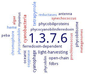
Word Map on EC 1.3.7.6
-
1.3.7.6
-
tetrapyrrole
-
cyanobacteria
-
light-harvesting
-
cyanophage
-
phycobiliproteins
-
heme
-
oceanic
-
phycobilisome
-
chromophore
-
fdbrs
-
ferredoxin-dependent
-
open-chain
-
synechococcus
-
myovirus
-
phycobilin
-
antenna
-
metagenomes
-
phycocyanobilin:ferredoxin
-
oxygenase
-
peba
-
prochlorococcus
-
reductases
-
phycocyanobilin
-
algae
- 1.3.7.6
- tetrapyrrole
- cyanobacteria
-
light-harvesting
-
cyanophage
-
phycobiliproteins
- heme
-
oceanic
-
phycobilisome
- chromophore
-
fdbrs
-
ferredoxin-dependent
-
open-chain
- synechococcus
-
myovirus
-
phycobilin
-
antenna
- metagenomes
-
phycocyanobilin:ferredoxin
- oxygenase
-
peba
- prochlorococcus
- reductases
- phycocyanobilin
- algae
Reaction
Synonyms
EBK42635, FDBR, ferredoxin-dependent bilin reductase, PcyX, PEB synthase, PebA, PebB, PebS, PhiPcyX, phycoerythrobilin synthase
ECTree

Advanced search results
Crystallization
Crystallization on EC 1.3.7.6 - phycoerythrobilin synthase
Please wait a moment until all data is loaded. This message will disappear when all data is loaded.
 top
topCRYSTALLIZATION (Commentary) 

ORGANISM
UNIPROT
LITERATURE
structures of substrate complex solved at 1.8- and 2.1 A resolution and of the substrate-free form at 1.55 A resolution. The overall folding reveals an alpha/beta/alpha-sandwich with similarity to the structure of phycocyanobilin:ferredoxin oxidoreductase. The substrate-binding site is located between the central beta-sheet and C-terminal alpha-helices. The substrate binding pocket shows a high flexibility. The substrate is either in a planar porphyrin-like conformation or in a helical conformation and is coordinated by a conserved aspartate/asparagine pair from the beta-sheet side. From the alpha-helix side, a conserved highlyflexible aspartate/proline pair is involved in substrate binding and presumably catalysis
purified recombinant substrate free form of PhiPcyX, sitting drop vapour diffusion method, mixing of 100 nl of 10-16.5 mg/ml protein in 20 mM TES-KOH pH 7.5, and 20 mM KCl, with 100 nl of reservoir solution containing 0.1 M Tris-HCl, pH 8.5, 0.2 M trimethylamine N-oxide (TMAO), and 20% w/v PEG MME 2000, at 4 °C. Final crystals used for structure determination grow at 4 °C via hanging drop vapour diffusion with 0.001 ml of 10 mg/ml protein in 20 mM TES-KOH pH 7.5, and 20 mM KCl mixed with 0.001 ml of 0.1 M Tris-HCl, pH 8.5, 0.05 M, TMAO, and 15% w/v PEG MME 2000 as reservoir solution, X-ray diffraction structure determination and analysis at 2.2 A resolution
-