1.1.1.28: D-lactate dehydrogenase
This is an abbreviated version!
For detailed information about D-lactate dehydrogenase, go to the full flat file.
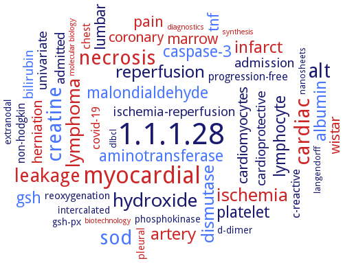
Word Map on EC 1.1.1.28
-
1.1.1.28
-
myocardial
-
creatine
-
cardiac
-
necrosis
-
alt
-
sod
-
leakage
-
lymphoma
-
hydroxide
-
ischemia
-
dismutase
-
reperfusion
-
albumin
-
infarct
-
artery
-
lymphocyte
-
aminotransferase
-
platelet
-
gsh
-
caspase-3
-
malondialdehyde
-
tnf
-
pain
-
lumbar
-
marrow
-
coronary
-
cardiomyocytes
-
herniation
-
wistar
-
univariate
-
admission
-
cardioprotective
-
bilirubin
-
admitted
-
ischemia-reperfusion
-
covid-19
-
c-reactive
-
non-hodgkin
-
chest
-
progression-free
-
pleural
-
reoxygenation
-
d-dimer
-
phosphokinase
-
extranodal
-
gsh-px
-
intercalated
-
dlbcl
-
nanosheets
-
langendorff
-
synthesis
-
molecular biology
-
diagnostics
-
biotechnology
- 1.1.1.28
- myocardial
- creatine
- cardiac
- necrosis
-
alt
- sod
- leakage
- lymphoma
-
hydroxide
- ischemia
- dismutase
-
reperfusion
- albumin
- infarct
- artery
- lymphocyte
- aminotransferase
- platelet
- gsh
- caspase-3
- malondialdehyde
- tnf
- pain
-
lumbar
- marrow
- coronary
- cardiomyocytes
- herniation
- wistar
-
univariate
-
admission
-
cardioprotective
- bilirubin
-
admitted
-
ischemia-reperfusion
- covid-19
-
c-reactive
-
non-hodgkin
- chest
-
progression-free
- pleural
-
reoxygenation
-
d-dimer
-
phosphokinase
-
extranodal
- gsh-px
-
intercalated
-
dlbcl
-
nanosheets
-
langendorff
- synthesis
- molecular biology
- diagnostics
- biotechnology
Reaction
Synonyms
D-(-)-lactate dehydrogenase, D-(-)-lactate dehydrogenase (NAD), D-isomer specific 2-hydroxyacid dehydrogenase NAD-binding protein, D-lactate dehydrogenase, D-lactic acid dehydrogenase, D-lactic dehydrogenase, D-LDH, D-LDH-like enzyme, D-LDH0653, D-LDH1, D-LDH2, D-LDH3, D-LDH82319, D-nLDH, D-specific lactic dehydrogenase, dehydrogenase, D-lactate, DLD1, DLDH, DLDH744, ECBD_2243, ECLDH, FD35_GL001981, Fermentative lactate dehydrogenase, FN0511, FNLDH, lactic acid dehydrogenase, ldb0101, Ldb1010, LDH, LdhA, ldhd, LDHD1, LDHD2, LDHD3, LdhTi, Ljd-LDH, MGG_01202, NAD-dependent D-lactate dehydrogenase, PA0927, PALDH, Respiratory D-lactate dehydrogenase, SO_0968, tp0037, WP_013906894
ECTree

Advanced search results
Crystallization
Crystallization on EC 1.1.1.28 - D-lactate dehydrogenase
Please wait a moment until all data is loaded. This message will disappear when all data is loaded.
 top
topCRYSTALLIZATION (Commentary) 

ORGANISM
UNIPROT
LITERATURE
purified recombinant enzyme, hanging drop vapour diffusion method, mixing of 0.002 ml of 15 mg/ml protein in 10 mM potassium phosphate, pH 7.0, with 0.002 ml of reservoir solution containing 14.4% PEG 8000, 80 mM cacodylate pH 6.5, 160 mM calcium acetate and 20% glycerol, and equilibration against 0.1 ml of reservoir solution, 25°C, 2 weeks, X-ray diffraction structure determination and analysis at 2.0 A resolution
purified recombinant enzyme in fully closed formation with lactate or pyruvate bound to the active site of each subunit of the functional dimer, 0.001 ml of protein solution containing 20.3 mg/ml protein in 20 mM Tris-HCl buffer pH 8.0 containing 150 mM NaCl, is mixed with 0.001 ml of reservoir solution containing 0.1 M MES buffer, pH 6.0, and 25% w/v PEG 200, 10 days, method optimization, X-ray diffraction structure determination and analysis at 2.12 A resolution, molecular replacement and structure modelling
hexagonal and tetragonal crystal forms, tetragonal form diffracted to 3.0 A resolution
-
comparison of the apo and ternary complex structures of Fusobacterium nucleatum FnLDH and Escherichia coli EcLDH and Pseudomonas aeruginosa PaLDH. FnLDH and EcLDH exhibit positive cooperativity in substrate binding, and PaLDH shows negatively cooperative substrate binding. The three enzymes consistently form homotetrameric structures with three symmetric axes, the P-, Q-, and R-axes, which allows apo-FnLDH and EcLDH to form wide intersubunit contact surfaces between the opened catalytic domains of the two Q-axis-related subunits in coordination with their asymmetric and distorted quaternary structures. apo-PaLDH possesses a highly symmetrical quaternary structure and partially closed catalytic domains that are favorable for initial substrate binding and forms virtually no intersubunit contact surface between the catalytic domains
crystals belong to the orthorhombic space group, diffract beyond 3.0 A resolution
-
purified recombinant selenomethionine-labeled enzyme, hanging drop vapour diffusion method, mixing of 0.0015 ml of 35 mg/ml protein in 40 mM Tris-HCl, pH 8.0, and 1 mM DTT with 0.0015 ml of reservoir solution containing 28% w/v PEG 400, 100 mM TrisHCl, pH 9.0, 200 mM magnesium sulfate at 22°C, X-ray diffraction structure determination and analysis at 2.0-2.1 A resolution by single wavelength anomalous dispersion using a selenomethionine derivative
-
molecular modeling and docking of substrates. D-LDH1 binds pyruvate using Tyr101, Arg235, and His296 by hydrogen bonds in the NADH-pyruvate-LDH1 complex
-
comparison of the apo and ternary complex structures of Fusobacterium nucleatum FnLDH and Escherichia coli EcLDH and Pseudomonas aeruginosa PaLDH. FnLDH and EcLDH exhibit positive cooperativity in substrate binding, and PaLDH shows negatively cooperative substrate binding. The three enzymes consistently form homotetrameric structures with three symmetric axes, the P-, Q-, and R-axes, which allows apo-FnLDH and EcLDH to form wide intersubunit contact surfaces between the opened catalytic domains of the two Q-axis-related subunits in coordination with their asymmetric and distorted quaternary structures. apo-PaLDH possesses a highly symmetrical quaternary structure and partially closed catalytic domains that are favorable for initial substrate binding and forms virtually no intersubunit contact surface between the catalytic domains
the complete tertiary structure of DLDH744 in complex with NAD+ is determined, PDB ID 4XKJ, determined by molecular replacement method and refined at 3.15 A-resolution
to 1.38 A resolution. The structure features two domains with a cleft between them. The smaller catalytic domain comprises a central beta-sheet flanked by alpha-helices. The sheet is composed of five parallel strands and one antiparallel one. The larger nucleotide-binding domain harbors a centrally located seven-strand parallel beta-sheet
comparison of the apo and ternary complex structures of Fusobacterium nucleatum FnLDH and Escherichia coli EcLDH and Pseudomonas aeruginosa PaLDH. FnLDH and EcLDH exhibit positive cooperativity in substrate binding, and PaLDH shows negatively cooperative substrate binding. The three enzymes consistently form homotetrameric structures with three symmetric axes, the P-, Q-, and R-axes, which allows apo-FnLDH and EcLDH to form wide intersubunit contact surfaces between the opened catalytic domains of the two Q-axis-related subunits in coordination with their asymmetric and distorted quaternary structures. apo-PaLDH possesses a highly symmetrical quaternary structure and partially closed catalytic domains that are favorable for initial substrate binding and forms virtually no intersubunit contact surface between the catalytic domains

comparison of the apo and ternary complex structures of Fusobacterium nucleatum FnLDH and Escherichia coli EcLDH and Pseudomonas aeruginosa PaLDH. FnLDH and EcLDH exhibit positive cooperativity in substrate binding, and PaLDH shows negatively cooperative substrate binding. The three enzymes consistently form homotetrameric structures with three symmetric axes, the P-, Q-, and R-axes, which allows apo-FnLDH and EcLDH to form wide intersubunit contact surfaces between the opened catalytic domains of the two Q-axis-related subunits in coordination with their asymmetric and distorted quaternary structures. apo-PaLDH possesses a highly symmetrical quaternary structure and partially closed catalytic domains that are favorable for initial substrate binding and forms virtually no intersubunit contact surface between the catalytic domains