1.1.1.2: alcohol dehydrogenase (NADP+)
This is an abbreviated version!
For detailed information about alcohol dehydrogenase (NADP+), go to the full flat file.
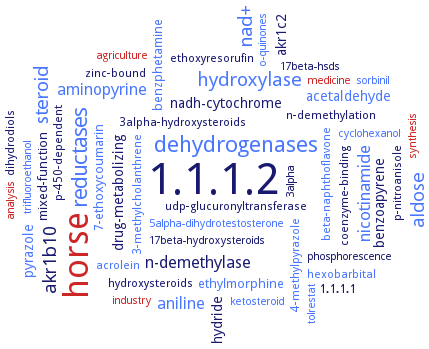
Word Map on EC 1.1.1.2
-
1.1.1.2
-
horse
-
dehydrogenases
-
reductases
-
hydroxylase
-
steroid
-
nad+
-
aldose
-
akr1b10
-
nicotinamide
-
aniline
-
aminopyrine
-
n-demethylase
-
akr1c2
-
acetaldehyde
-
pyrazole
-
nadh-cytochrome
-
drug-metabolizing
-
benzoapyrene
-
hydride
-
benzphetamine
-
ethylmorphine
-
mixed-function
-
1.1.1.1
-
7-ethoxycoumarin
-
beta-naphthoflavone
-
udp-glucuronyltransferase
-
p-nitroanisole
-
4-methylpyrazole
-
3alpha-hydroxysteroids
-
coenzyme-binding
-
dihydrodiols
-
3-methylcholanthrene
-
n-demethylation
-
ethoxyresorufin
-
hydroxysteroids
-
acrolein
-
hexobarbital
-
p-450-dependent
-
zinc-bound
-
17beta-hsds
-
analysis
-
sorbinil
-
cyclohexanol
-
medicine
-
agriculture
-
trifluoroethanol
-
5alpha-dihydrotestosterone
-
ketosteroid
-
synthesis
-
17beta-hydroxysteroids
-
industry
-
o-quinones
-
tolrestat
-
3alpha
-
phosphorescence
- 1.1.1.2
- horse
- dehydrogenases
- reductases
- hydroxylase
- steroid
- nad+
- aldose
- akr1b10
- nicotinamide
- aniline
- aminopyrine
- n-demethylase
- akr1c2
- acetaldehyde
- pyrazole
-
nadh-cytochrome
-
drug-metabolizing
-
benzoapyrene
-
hydride
- benzphetamine
- ethylmorphine
-
mixed-function
-
1.1.1.1
- 7-ethoxycoumarin
- beta-naphthoflavone
-
udp-glucuronyltransferase
-
p-nitroanisole
- 4-methylpyrazole
-
3alpha-hydroxysteroids
-
coenzyme-binding
-
dihydrodiols
- 3-methylcholanthrene
-
n-demethylation
-
ethoxyresorufin
- hydroxysteroids
- acrolein
- hexobarbital
-
p-450-dependent
-
zinc-bound
- 17beta-hsds
- analysis
- sorbinil
- cyclohexanol
- medicine
- agriculture
- trifluoroethanol
- 5alpha-dihydrotestosterone
- ketosteroid
- synthesis
-
17beta-hydroxysteroids
- industry
- o-quinones
- tolrestat
-
3alpha
-
phosphorescence
Reaction
Synonyms
2� Adh, 3-DG-reducing enzyme, A3L14_07690, AAur_2040, ADH, ADH-1, ADH-2, ADH-I, ADH-II, ADH2, Adh319, ADH4, Adh6, AdhA, AdhB, AdhD, AKR1A1, AKR1A4, alcohol dehydrogenase C, alcohol dehydrogenase D, alcohol dehydrogenase [NADP(+)], Alcohol dehydrogenase [NADP+], aldehyde reductase, aldehyde reductase (NADPH2), aldehyde/ketone reductase, aldo-keto reductase, Aldo-keto reductase family 1 member A1, aldose reductase, ALDR, alipathic aldehyde reductase, ALR, ALR 1, ALR1, BdhA, bovine brain aldehyde reductase, CbADH, D-glucuronate reductase, daunorubicin reductase, DRD, EhADH1, FeADH, Gre2p, hexogenate dehydrogenase, high-Km aldehyde reductase, HvADH2, iron-containing alcohol dehydrogenase, KpADH, L-hexonate dehydrogenase, LB-RADH, LBADH, liver alcohol dehydrogenase, low-Km aldehyde reductase, mevaldate reductase, Mpd1, NADP(H)-dependent alcohol dehydrogenase, NADP+-dependent alcohol dehydrogenase, NADP-alcohol dehydrogenase, NADP-aldehyde reductase, NADP-dependent aldehyde reductase, NADP-linked aryl alcohol dehydrogenase, NADPH-aldehyde reductase, NADPH-cytochrome c reductase, NADPH-dependent ADH, NADPH-dependent aldehyde reductase, NADPH-dependent FALDR, NADPH-dependent fatty aldehyde reductase, NADPH-linked aldehyde reductase, nonspecific succinic semialdehyde reductase, Octopine dehydrogenase, putative iron alcohol dehydrogenase, PyAeADHII, rabbit kidney aldehyde-ketone reductase, RADH, ScADHVI, short-chain ADH, short-chain alcohol dehydrogenase, short-chain dehydrogenase/reductase, TBADH, TbADH1, Teth39_1597, Teth514_0564, TPN-L-hexonate dehydrogenase, TPNH-linked aldehyde reductase, TPNH-specific aldehyde reductase, triphosphopyridine nucleotide-linked aldehyde reductase, TsAdh319, Y63 protein, yeast alcohol dehydrogenase, YqhD
ECTree

Advanced search results
Crystallization
Crystallization on EC 1.1.1.2 - alcohol dehydrogenase (NADP+)
Please wait a moment until all data is loaded. This message will disappear when all data is loaded.
 top
topCRYSTALLIZATION (Commentary) 

ORGANISM
UNIPROT
LITERATURE
crystals of D275PEhADH1 are grown using the hanging-drop vapor-diffusion method at 20°C. Crystal structure of the thermostabilized mutant D275P-EhADH1 suggests that a proline residue at position 275 thermostabilizes the enzymes by reducing flexibility and by reinforcing hydrophobic interactions at the dimerdimer interface of the tetrameric ADH
16°C, wild-type RADH: 15 mg/ml stock solution (21% polyethyleneglycol monomethyl ether 550, 0.1 M Tris-HCl, 50 nM MgCl2, 40 mM acetophenone, 10 mM NADP+), growth for 3 weeks, max. size 0.25 mm x 0.25 mm x 1.0 mm, RADH-G37D: 21 mg/ml stock solution (methyl-2,4-pentanediol, 20% polyethyleneglycol 400 or polyethyleneglycol monomethyl ether 550, 0.1 M Hepes, 50 mM MgCl2 59 mM 1-phenylethanol, 25 mM NADH), growth 1 week, max. size 0.4 mm x 0.5 mm x 1.7 mm
-
in presence and absence of NADP+, structural features of an independent ADH class
-
sitting drop vapour diffusion plates at 20°C. Crystal structure and X-ray fluorescence of wild-type and cobalt-substituted enzyme. Cocrystal structure of the enzyme with NADPH and analysis of the putative active site
in complex with NADP+, to 3.2 A resolution. Crystals belong to space group P21
-
at 22°C by vapor-diffusion using the hanging drop method. ALR1 in ternary complex with the coenzyme NADPH and 3,5-dichlorosalicylic acid, at a resolution of 2.41 A
hanging drop method, with NADPH, ammonium sulfate, Tris HCl-buffer, pH 8.1, buffer C, maximum side: 0.3 mm x 0.1 mm x 0,1 mm after 1 week
-
in ternary complex with NADPH and a 5-arylidene-2,4-thiazolidinedione aldose reductase inhibitor, [5-(3-carboxymethoxy-4-methoxybenzylidene)-2,4-dioxothiazolidin-3-yl]acetic acid to 1.99 A resolution. The partially disordered inhibitor forms a tight network of hydrogen bonds with the active site residues Tyr50 and His113 and coenzyme. pi-Stacking interactions with several conserved active site tryptophan residues and hydrogen-bonding interactions with the non-conserved C-terminal residue Pro301 in aldehyde reductase ALR1 contribute to inhibitor selectivity. In particular for the potent inhibitor [5-(3-carboxymethoxy-4-methoxybenzylidene)-2,4-dioxothiazolidin-3-yl]acetic acid, the rotameric state of the conserved residue Trp220 in ALR1, i.e Trp 219 in aldose reductase, is important in forming a pi-stacking interaction with the inhibitor in aldose reductase and contributes to the difference in the binding of the inhibitor to the enzymes
purified aldehyde reductase, ALR1, in ternary complex with NADPH and [5-(3-carboxymethoxy-4-methoxybenzylidene)-2,4-dioxothiazolidin-3-yl]acetic acid, hanging drop method, 17-18 mg/ml protein in 5 mM Tris-HCl, pH 6.5, containing 5 mM 2-mercaptoethanol, mixed with NADPH and inhibitor in a 1:20:3molar ratio, the reservoir solution contains 2.0 M ammonium sulfate, and 0.1 M Tris-HCl buffer, pH 8.5, 10 days, X-ray diffraction structure determination and analysis at 1.99 A resolution
crystals of P275DTbADH1 are grown using the hanging-drop vapor-diffusion method at 20°C. Crystal structure of the thermostabilized mutant P275D-EhADH1 suggests that a proline residue at position 275 thermostabilizes the enzymes by reducing flexibility and by reinforcing hydrophobic interactions at the dimerdimer interface of the tetrameric ADH
crystal structure of the enzyme in a binary complex with 5-hydroxy-NADP at 1.68 A resolution
to 1.68 A resolution, crystals belong to space group I222, with unit-cell parameters a = 55.63, b = 83.25, c = 120.75 A
purified enzyme in unit cells belonging to space groups P21, P212121 and P43212 (monoclinic, orthorhombic, and tetragonal crystals), vapour diffusion method, mixing of 0.0012 ml of 10 mg/ml protein in 50 mM Tris, pH 7.5, and 50 mM NaCl, with 600 nl of reservoir solution containing 8% PEG 4000, pH 4.2-4.5, in a protein:reservoir ratio of 2:1 (1:1 for the tetragonal crystals), at 20°C, X-ray diffraction structure determination and analysis at 2.4 A, 2.1 A, and 1.9 A resolution, respectively, modeling by molecular replacement using the FeADH from Thermotoga maritima as a template